

National Antivisection Society
Researchers create reliable complex model of human breast cancer
27 January 2011
Breast cancer is a complex and heterogeneous disease with several different molecular alterations involved in its pathogenesis and progression.
A comprehensive approach that takes into account the complexity of the disease is required to improve the efficacy of target-based therapy in breast cancer. None of the above can be easily or accurately replicated in animal models.
Currently, the two types of animal model commonly used to study breast cancer are transgenic mice and xenograft models. These models do not allow an understanding of the biology of the tumour, nor do they take into account the multiple human cell types involved in breast cancer development and progression. To define cancer progression animal models use a number of end points in their assays including tumour cell proliferation, apoptosis (cell death) and release of matrix degrading enzymes.
The all human model
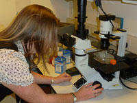
Dr Debbie Holliday, and her team have developed a novel 3D model of breast cancer. This model is the first to contain the 3 major epithelial (lining) and stromal (connective and supporting) components of the breast; luminal epithelial, myoepithelial and fibroblasts. When validated against human tumours the morphological appearance bore a remarkable resemblance to human breast tumours.With this model the team at the Leeds Institute of Molecular Medicine, based in St James’s University Hospital, have shown the ability of human tumour associated stromal fibroblasts to disrupt tumour structure formation. They also found that this process can be inhibited by the presence of drugs. The 3D model will be derived from ERα+ breast cancer, which represents two-thirds of all breast cancers. The samples have been obtained from consenting patients.
The primary aim of this project to is validate two novel in vitro models of breast cancer (the 3D model and the slice) against published endpoints of cancer progression using published data on animal experiments. This will demonstrate that the models provide data comparable with those observed in animal experiments, thus showing that animal models can be replaced, by the more superior and relevant human model.
In addition, it will be necessary to validate that the 3D culture model maintains a phenotype which is representative of the original tumour. To do this the researchers will grow isolated cells from primary tumours in their 3D culture model and compare tissue slices of these cultures to samples from the same case. With this the study will be producing a valid method for testing tumour response to therapies. Indeed, the effect of 3 commonly used breast cancer therapies will be assessed in the unique model systems by including them in the culture medium for 7 days. Slices of the cultured tissue can then be fixed and analysed and the endpoints, commonly used in animal models of cancer progression, will be measured in the models.
The future of cancer research
The unique strength of this model is that it contains not only human tumour cells but incorporates a human only stromal component, something which is not achievable in animal models. With this well defined, highly reproducible model it is possible to genetically modify individual cell populations to look at the effect either in isolation or in combination with other factors. This in vitro system also provides a tool to investigate potential new therapeutic agents.Dr Valerie Speirs, who works alongside Dr Holliday, has recently expanded this tissue slice research to encompass pancreatic cancer to explore its potential for adenoviral delivery as a novel therapeutic approach.

